Radiological tests for Morton syndrome
The examinations are of little use in the typical forms of Morton’s nevroma, for which the diagnosis must remain clinical. Nevertheless in our modern age, it is necessary to confirm the suspicion of Morton’s syndrome, before any therapeutic management. The practice of standard x-rays eliminates an associated pathology or other.
In case of doubt or entanglement with other pathologies of the forefoot, the assessment is completed by practice:
- an ultrasound, an easy access exam but that is still a “descrambling” exam and dependent on the radiologist’s experience,
- an MRI scan.
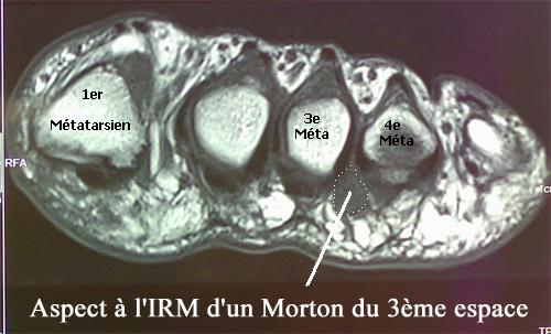
The MRI is the reference study, which makes it possible to diagnose Morton’s neuroma easily, but also to locate it precisely.
Normal ultrasound and MRI do not eliminate the diagnosis. Sometimes it is necessary to repeat the exam remotely until the swelling is visible…
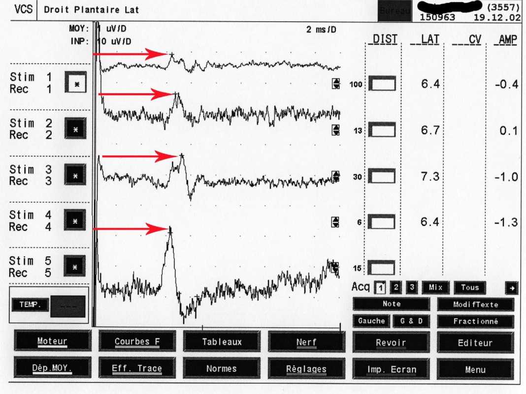
Finally, in unusual forms of Morton syndrome, the realization of an Electromyogram can be useful. The neurologist using specific stimulation electrodes will study nerve conduction rates, and amplitudes of sensitive potentials. The examination may be in favor of the diagnosis of Morton syndrome, but also will rule out other possible diagnoses.


